
(© MST – stock.adobe.com)
IVF advancement: New camera techniques transform biological imaging
In a nutshell
- Optimizing camera settings can provide better images of embryos than increasing light intensity, which protects delicate specimens from damage
- Techniques like pixel binning (combining pixels) and longer exposure times increased image quality by 3x without additional light
- These quantum-inspired camera methods could revolutionize biological imaging, especially for light-sensitive specimens like those used in IVF research
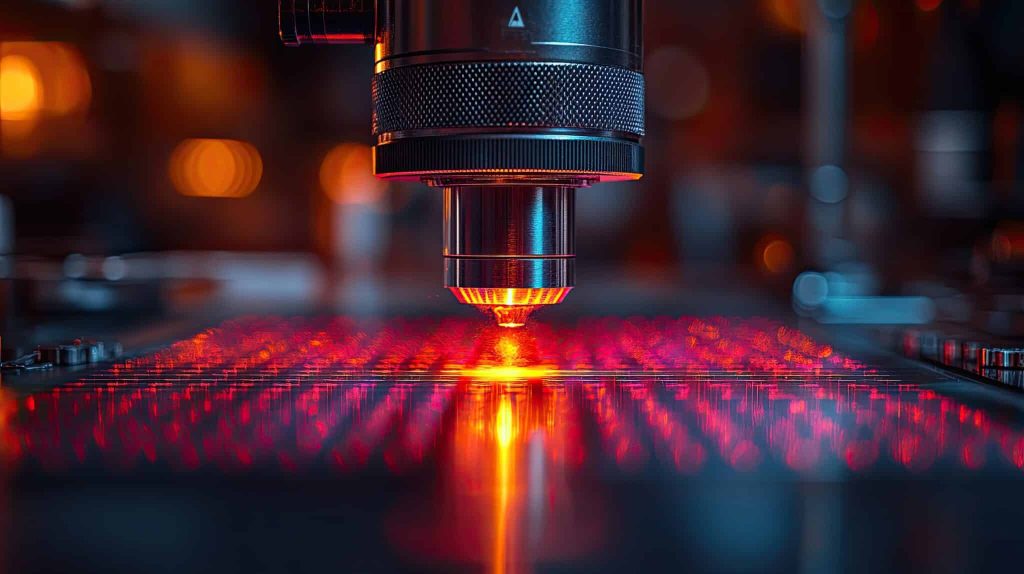
ADELAIDE, Australia — In a breakthrough at the intersection of quantum physics and biology, researchers at the University of Adelaide have figured out a way to observe the earliest stages of life. Using cameras designed for quantum measurements, these scientists have captured unprecedented images of developing embryos while minimizing the damage typically caused by conventional imaging techniques.
Their research, published in the journal APL Photonics, serves as a roadmap for scientists navigating the world of low-light imaging. The central idea is both simple and powerful: by optimizing camera settings and employing quantum-inspired detection technology rather than increasing illumination intensity, researchers can achieve better image quality while minimizing damage to living specimens.
“Damage from illumination is a real concern which can often be overlooked. Using the lowest level of light possible, together with these very sensitive cameras is important for understanding biology in live and developing cells,” says Professor Kishan Dholakia, director of the University’s Centre of Light for Life and one of the study’s authors, in a statement.
Consider the delicate mouse embryo, a living structure so sensitive to light that excessive illumination can disrupt its development or even kill it. Yet scientists need clear images to study its intricate cellular processes. This is the fundamental challenge of biological imaging—getting enough signal without destroying the very thing you’re trying to observe.
“These samples are living, developing specimens that serve as a foundation for studies supporting advancements in clinical IVF,” notes Professor Dholakia.

Beyond the camera: Why the right settings are crucial
The research shows that picking the right camera is just the first step. What really matters is knowing how to optimize settings for specific imaging challenges. By testing three different camera types—the ORCA-Flash V3, ORCA-Quest, and iXON Life 888—the team demonstrated how tweaking various settings dramatically improved image quality without increasing light intensity.
One key setting is “pixel binning,” which combines multiple pixels into one larger virtual pixel. This allows the camera to collect more light in each measurement, though at the cost of some image detail. Another important setting is exposure time—how long the camera collects light before taking a reading.
In one striking example, when imaging a mouse embryo, switching from standard settings to optimized settings (2×2 pixel binning, longer exposure time, and specialized camera mode) boosted the signal-to-noise ratio from 1.78 to 5.6—without any additional illumination. Signal-to-noise ratio (SNR) is a measure of how clear and reliable an image is—higher numbers mean cleaner images where the biological structures stand out more clearly from background noise.
The science behind these improvements relates to light’s fundamental nature. At extremely low intensities, light behaves more like individual particles (photons) than waves. Modern quantum-inspired cameras can detect these individual packets of light energy at each pixel, allowing for unprecedented sensitivity.
“A lot of natural compounds in cells light up when illuminated, and this can tell us a lot about what we’re looking at, but unfortunately the signal is very weak,” said lead author and PhD student Zane Peterkovic.
The differences between cameras
Different camera designs handle this challenge in different ways. sCMOS (scientific Complementary Metal Oxide Semiconductor) cameras like the ORCA-Flash and ORCA-Quest offer excellent resolution with relatively low noise, working similarly to advanced versions of digital camera sensors in smartphones but with much higher precision. Meanwhile, EMCCD (Electron-Multiplying Charge-Coupled Device) cameras like the iXON Life excel at detecting extremely faint signals by amplifying electrons before they’re read out—similar to turning up the volume on a weak audio signal before processing it.
Each camera type has its trade-offs. EMCCDs are incredibly sensitive but suffer from “excess noise factor”—the amplification process itself introduces additional uncertainty, like how turning up the volume also amplifies static in an audio recording. sCMOS cameras have lower sensitivity but don’t have this excess noise problem.

The paper avoids getting stuck in theory, focusing instead on practical examples. When imaging embryos that naturally emit light without added dyes, the researchers show how to balance pixel size, exposure time, and camera mode for best results.
“A large part of the project involved developing a method to fairly compare the image quality across different cameras,” explains Peterkovic.
Perfecting the image
One key finding challenges a common practice. Many scientists instinctively crank up magnification to capture more detail, but the research shows there’s a physical limit to how much detail can be resolved due to light diffraction. Higher magnification beyond this point just spreads the same information across more pixels, reducing signal intensity without gaining additional information. This insight helps researchers select appropriate magnification that maximizes signal without unnecessary light exposure.
The team also tested machine learning algorithms to enhance low-light images. Three different denoising algorithms—Noise2Fast, Neighbor2Neighbor, and Accurate Correction of sCMOS Noise (ACsN)—showed promise but came with warnings about potential artifacts or “hallucinations” if not used carefully. Denoising algorithms work like digital filters that try to remove random speckles and static from images while preserving the true biological structures.
“We even explored how AI can be used to remove noise from the captured images, which is essentially static because the camera struggles to capture enough light,” says Peterkovic. “These steps go beyond just putting the camera in the microscope to take pictures.”

The excitement of quantum camera tech
For scientists working with living specimens, the benefits are clear. These optimization strategies reduce phototoxicity and photobleaching—two major problems in live-cell imaging. Phototoxicity refers to how light can poison or damage living cells, similar to severe sunburn at a cellular level. Photobleaching is when fluorescent molecules fade under light exposure, like how fabrics fade in strong sunlight. Both problems are reduced when less light is needed for imaging.
“It’s exciting to apply these quantum cameras and use it to get the most out of our microscopes,” Peterkovic adds. “Digital camera technology has advanced to the point where fundamental physics concepts like quantum mechanics become important and relevant.”
Surprisingly, the tutorial reveals that even relatively inexpensive cameras can achieve impressive results when properly optimized, making high-quality imaging more accessible to labs with limited resources.
“Modern imaging technology is very exciting with what it enables us to see,” remarked Professor Dholakia.
The authors argue that understanding camera physics and optimization techniques isn’t merely a technical exercise—it’s essential for responsible research that minimizes harm to living specimens while maximizing scientific insight. As microscopy pushes the boundaries of what we can see in the biological world, these principles will only grow in importance.
Future work will extend into quantum imaging, where quantum states of light may reveal even more information about samples. For low-light microscopy, the message is clear: quantum-inspired cameras that detect the faintest signals can revolutionize how we study the beginning of life.
Paper Summary
Methodology
The researchers evaluated camera performance and optimization strategies through several steps. First, they characterized the noise patterns of three different scientific cameras: two scientific CMOS cameras (ORCA-Flash V3 and ORCA-Quest) and one electron-multiplying CCD camera (iXON Life 888). This involved creating charts that show how noise changes with signal intensity and generating maps that identify patterns in camera noise. To test these cameras in real situations, they built a specialized microscope called a two-photon light sheet fluorescence microscope, which gently images living specimens by illuminating only a thin plane of the sample at a time. They used live mouse embryos at the blastocyst stage (about 4-5 days post-fertilization) as test subjects because these embryos naturally emit light from certain molecules, making them ideal for low-light imaging tests. The researchers then systematically tested how different camera settings—combining adjacent pixels (called “binning”), exposure time, specialized operating modes, and magnification—affected image quality. They measured image quality using signal-to-noise ratio (SNR) and contrast-to-noise ratio (CNR), with a custom program that objectively calculates these values from sets of identical images. Finally, they tested three different AI-based denoising algorithms to see how well they could enhance low-light images.
Results
The results showed several key insights for getting better low-light images. First, the researchers found that pixel size greatly affects image quality—larger pixels collect more light, improving signal-to-noise ratio. For example, combining 2×2 pixels (called “binning”) nearly doubled the SNR in some cases, though this reduces the detail in the image. Second, using longer exposure times improved image quality, though with diminishing returns as background noise was also captured for longer periods. Third, specialized camera modes showed significant benefits: the “light sheet mode” of sCMOS cameras, which synchronizes row reading with light sheet scanning, dramatically reduced background noise, while the “photon resolving mode” of the ORCA-Quest reduced electronic noise to enable single photon detection. When comparing camera types, the EMCCD camera (iXON Life) performed better for extremely dim samples with minimal background, but sCMOS cameras worked better when background fluorescence was significant. Most importantly, optimized camera settings (2×2 pixel binning, longer exposure time, specialized operating mode) achieved better results than simply doubling the light intensity, with SNR improvements from 1.78 to 5.6 compared to just 2.47 with increased illumination. Testing of AI denoising algorithms showed mixed results, with some algorithms significantly improving image quality while others introduced artifacts or false features.
Limitations
The researchers acknowledge several limitations to their study. First, they only tested three specific cameras, which might not represent all camera technologies available for microscopy. Second, the experiments used a specific microscopy technique and mouse embryos as samples, so the findings may not apply perfectly to all microscopy setups or biological specimens. The AI denoising algorithms were evaluated on a limited dataset, and newer algorithms might offer better performance as this field rapidly advances. Additionally, their optimization strategies involve trade-offs—better signal often comes at the cost of detail or time resolution, which may not work for all experimental needs. The analysis also didn’t extensively evaluate very high-speed imaging applications or specialized techniques requiring precise timing. Finally, while the tutorial provides guidance on camera selection and optimization, it emphasizes that each imaging challenge requires its own specific approach, and there is no one-solution-fits-all for low-light microscopy.
Discussion and Takeaways
The most significant lesson from this study is that thoughtful optimization of camera settings can often achieve better results than simply increasing light intensity, which can damage delicate biological specimens. The researchers emphasize that understanding how cameras work—particularly how different sources of noise affect image quality—is essential for quantitative microscopy. They highlight the importance of matching pixel size to the optical resolution of the microscope, noting that using more pixels than necessary to capture the available resolution reduces signal intensity without gaining additional information. Another key insight is that different camera designs have distinct advantages: EMCCD cameras excel in extremely low-light conditions with minimal background, while sCMOS cameras perform better when background fluorescence is significant. The tutorial also provides practical guidance on evaluating image quality objectively using signal-to-noise ratio (SNR) and contrast-to-noise ratio (CNR) rather than relying on subjective visual assessment. For AI approaches to image enhancement, the researchers caution that while denoising algorithms can improve image quality, they should be used carefully and only after optimizing physical imaging parameters. Finally, the tutorial emphasizes that image optimization depends heavily on the specific experimental goals—whether prioritizing detail, speed, or minimal light exposure—and researchers should tailor their approach accordingly.
Funding and Disclosures
The research was supported by several funding sources, including the Australian Research Council (ARC Grant No. FL210100099), the National Health and Medical Research Council (NHMRC, Grant No. APP2003786), and the Australian Research Council Centre of Excellence in Optical Microcombs for Breakthrough Science (Project No. CE230100006). The authors declared no conflicts of interest related to this work. They acknowledged the loan of an iXON Life 888 camera from Paul Wardill and Michael Buckett of Coherent Scientific Ltd., which allowed them to compare different camera technologies.
Publication Information
The tutorial titled “Optimizing image capture for low-light widefield quantitative fluorescence microscopy” was published in APL Photonics (Volume 10, Issue 031102) on March 12, 2025. The paper, authored by Zane Peterkovic, Avinash Upadhya, Christopher Perrella, Admir Bajraktarevic, Ramses E. Bautista Gonzalez, Megan Lim, Kylie R. Dunning, and Kishan Dholakia, represents a collaboration between researchers at the Centre of Light for Life and School of Biological Sciences at the University of Adelaide in Australia, the Centre of Light for Life and The Robinson Research Institute at the University of Adelaide, and the School of Physics and Astronomy at the University of St Andrews in the United Kingdom. The paper can be accessed online at https://doi.org/10.1063/5.0245239.