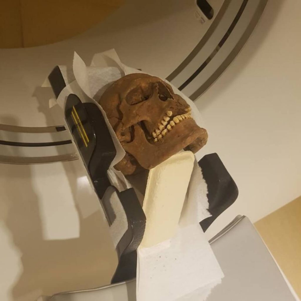

The skulls of Viking-era individuals were examined with modern computed tomography, in the search for infections, inflammations and other diseases. (Credit:
Photo by Carolina Bertilsson)
In a nutshell
- CT scans of 15 Viking-age skulls revealed widespread dental infections, sinusitis, ear diseases, and joint problems that would have caused significant pain and reduced quality of life in a time before modern medicine.
- 80% of the examined individuals showed evidence of tooth root infections, with many cases so severe that the infection had broken through bone into the sinus cavity or oral cavity.
- The study demonstrates how modern medical imaging technology can reveal health conditions in archaeological remains that would be impossible to detect through traditional examination methods, allowing researchers to preserve specimens while still extracting valuable information.
GOTHENBURG, Sweden — When archaeologists unearthed the remains of Viking-age Swedes from their thousand-year slumber, they expected to find evidence of battle wounds and hard living. Instead, using cutting-edge medical technology, they discovered skeletal evidence suggesting something far more relatable—a population suffering from toothaches, sinus infections, and ear problems. A new study from the University of Gothenburg reveals that despite their legendary status, Vikings endured many of the same painful health conditions modern humans face, but without the benefit of antibiotics, pain relievers, or medical intervention.
The study, published in BDJ Open, examined the remains of 15 Viking-era individuals from Varnhem, Sweden, dating back to the 10th-12th century A.D. Using computed tomography (CT) scans, the same technology that creates detailed 3D images in modern hospitals, the researchers were able to see inside these ancient skulls without damaging the precious remains.
For anyone who romanticizes life in the Viking age as a time of legendary warriors and epic sagas, this research offers a stark reminder of the harsh realities faced by people living without modern medicine. The scans revealed widespread dental problems, including cavities, gum disease, and tooth infections that had eaten through the jawbone, conditions that would have caused significant pain and difficulty eating.
More surprisingly, they discovered evidence of chronic sinusitis, middle ear infections, and other inflammatory conditions that would have been impossible to detect through traditional archaeological examination. These hidden ailments suggest that many Viking-age individuals suffered from persistent health problems that might never have been documented without this technology.

Photo by Carolina Bertilsson)
These findings indicate that the population suffered from numerous orofacial pathologies, including dental disease, sinusitis, otitis, and various infections. In an era before antibiotics or modern dental care, such conditions could become serious, even life-threatening.
The skulls examined in this study came from one of Sweden’s earliest Christian settlements. Excavated in 2005 from the grounds behind Varnhem Abbey, these remains represent a transitional period in Scandinavian history, when the region was moving from traditional Norse beliefs toward Christianity. Varnhem is known for its thousands of ancient graves and excavations of well-preserved skeletons, making it an ideal site for such research.
“There was much to look at. We found many signs of disease in these individuals. Exactly why we don’t know. While we can’t study the damage in the soft tissue because it’s no longer there, we can see the traces left in the skeletal structures,” says lead study author Carolina Bertilsson, an assistant researcher at the University of Gothenburg and a dentist within Sweden’s Public Dental Service, in a statement.
About a year before this investigation, researchers published findings based on examining teeth from the same Viking Age population of Varnhem. The current study takes that research further by examining entire skulls, not just teeth, using sophisticated CT technology.

This approach is revolutionary because it reveals health conditions that would be virtually impossible to detect through standard archaeological examination. Traditional methods involve examining bones under strong light, sometimes using dental tools for closer inspection. But this approach can’t see inside intact bone or closed skull chambers.
CT scanning changes everything. By creating cross-sectional images of the skull, the technology allows researchers to examine internal structures without physically cutting into the specimens. This is crucial for preserving irreplaceable archaeological materials while still extracting valuable information from them. The three-dimensional images enable researchers to study in detail the various types of skeletal damage, layer by layer, in different parts of the skull.
Among the most common findings were signs of periodontal disease, what we today would call severe gum disease. Ten of the 15 individuals showed evidence of this condition, which causes the bone supporting the teeth to deteriorate. The researchers noted that this was especially prevalent in the back molars, though some individuals had bone loss around their front teeth as well.
Even more prevalent were signs of periapical inflammatory disease, essentially infections at the roots of teeth. A startling 80% of the examined individuals (12 out of 15) showed evidence of these infections. In some cases, these infections had eaten through the bone, creating pathways into the maxillary sinus or the oral cavity, conditions that would have been extremely painful and potentially dangerous.
One individual, identified as number 12 in the study, had a cyst-like lesion around tooth 36 (a lower left molar). This same individual also showed signs of sclerotization (hardening) of the mastoid process, a part of the skull behind the ear. In modern medicine, this finding is associated with acute or chronic middle ear infections, which, without antibiotics, could spread and potentially become fatal.

Photo by Paula Morad)
The temporomandibular joint (TMJ), the hinge that connects the jaw to the skull, also showed frequent abnormalities. Over half of the studied individuals had TMJ problems, including bone spurs, flattening of joint surfaces, and erosion. These conditions would have caused pain when chewing and possibly limited jaw movement. Anyone who has experienced jaw pain knows how debilitating it can be, affecting basic daily activities like eating and speaking.
Several individuals also showed signs of chronic sinusitis. The researchers identified thickened bone around the sinus cavities in 20% of the skulls, indicating long-term inflammation. Anyone who has experienced sinus infections can relate to the discomfort these Viking-age people would have endured, such as nasal congestion, facial pressure, and reduced sense of smell, but without the benefit of antibiotics or decongestants.
One individual (number 98) showed evidence of periosteal bone formation. This is essentially a new bone growing on top of existing bone in response to inflammation. This reaction can be triggered by various conditions, including trauma, arthritis, and infections. In a world without painkillers or anti-inflammatory medications, such conditions would have caused ongoing discomfort and limited mobility.
The researchers also found unusual anatomical variations in some individuals. One person had a three-rooted premolar, and another had bony growths on the roof of their mouth called tori palatini. While not necessarily pathological, these variations add to our understanding of the biological diversity within this population.
“The results of the study provide greater understanding of these people’s health and wellbeing. Everyone knows what it’s like to have pain somewhere, you can get quite desperate for help. But back then, they didn’t have the medical and dental care we do, or the kind of pain relief – and antibiotics – we now have. If you developed an infection, it could stick around for a long time,” explains Bertilsson.
The researchers were careful to distinguish between damage that occurred during life (ante-mortem) and changes that happened after death (post-mortem). They only reported conditions that showed characteristics of processes that would have occurred while the person was alive. This methodical approach ensures that the health conditions they identified were actually experienced by these individuals during their lifetimes.
While this study focused on just 15 individuals from a specific place and time, it provides a compelling glimpse into the health challenges faced by people during the Viking era. The prevalence of dental and sinus problems suggests a population dealing with chronic pain and infection as part of everyday life. All the skulls came from adults who died between 20 and 60 years of age, representing a cross-section of the adult Viking population in this settlement.
For archaeologists and medical historians, this research demonstrates the value of modern imaging technology in understanding ancient populations. For the rest of us, it serves as a reminder of how far medical science has progressed. Many of the conditions that plagued these Viking-age individuals are easily treatable today with antibiotics and other modern interventions.
Most importantly, this research helps us see past the mythologized version of Vikings that dominates popular culture, revealing instead real humans who struggled with the same types of health problems that affect people today. We may imagine them as fearless warriors sailing the seas, but the reality included individuals suffering from painful infections, reminders of our shared human vulnerability across the centuries.
“Very many of today’s archaeological methods are invasive, with the need to remove bone or other tissue for analysis. This way, we can keep the remains completely intact yet still extract a great deal of information,” says Bertilsson.
As we continue to develop and apply new technologies to archaeological remains, we can gain even more insights into the lives of our ancestors. For now, the next time you take an antibiotic for a sinus infection or visit a dentist for a cavity, consider the Viking-age individuals who faced these same ailments without modern medicine—a stark reminder of how medical advances have transformed the human experience.
Paper Summary
Methodology
The researchers selected 15 skulls from a larger collection of 171 Viking-age individuals that had previously undergone dental examination. The selection was based on convenience sampling, with the main criteria being that the skull had to include the cranium (with or without the lower jaw) and be possible to mount in the CT scanner. The sample consisted of nine males and six females, with estimated ages at death ranging from 24 to 60 years. The CT examination was performed using an Optima CT660 scanner, which created thin slice images (0.625 mm thickness) that were then exported to a medical imaging system for detailed analysis. Three dental professionals—two specialists in oral and maxillofacial radiology with forensic experience and one general dentist with archaeological experience—examined the images together to identify pathological conditions. They carefully distinguished between conditions that occurred during life and changes that happened after death, focusing only on the former for their analysis. This non-invasive approach allowed them to examine internal structures of the skulls without damaging the valuable archaeological specimens.
Results
The CT scans revealed a high prevalence of dental and oral health issues among the Viking-age population. Ten of the 15 individuals showed evidence of periodontal disease, characterized by horizontal bone loss, vertical bone defects, and involvement of the furcation (the area where tooth roots divide). Even more striking was that 12 of the 15 individuals (80%) had signs of periapical inflammatory disease—essentially, infections at the tooth roots that in some cases had broken through the bone into the sinus cavity or oral cavity. Over half of the studied individuals (8 of 15) showed abnormalities of the temporomandibular joint, including bone spurs, flattening of joint surfaces, and erosion. Other findings included dental cavities in 4 individuals, retained tooth roots, bone destruction, and evidence of chronic sinusitis in 3 individuals. One individual showed sclerotization of the mastoid process, suggesting middle ear infection, while another had bone destruction near the ear canal. These findings paint a picture of a population dealing with numerous painful oral and facial conditions that would have significantly impacted quality of life.
Limitations
The researchers acknowledge several limitations to their study. First and foremost is the small sample size—only 15 individuals from a much larger population. This means the findings cannot be generalized to the entire Viking population. There was also an uneven distribution between males (9) and females (6), making it impossible to draw conclusions about sex differences in health conditions. Many potential study subjects had to be excluded because their remains were too fragmented to be examined with CT scanning. The researchers also note that post-mortem tooth loss represents a potential source of bias, particularly when trying to determine dental pathologies. Additionally, the transportation of skulls to the CT facility might have caused some post-mortem alterations, and in several individuals, debris trapped in skull cavities made interpretation of some areas difficult. Despite these limitations, the study provides valuable insights into health conditions that would be impossible to detect through traditional archaeological methods.
Discussion and Takeaways
This exploratory study demonstrates the value of CT scanning as a complementary method for examining archaeological remains. While some conditions identified in the study could have been detected through traditional examination, the CT scans revealed important findings that would otherwise have remained hidden—particularly conditions affecting internal structures like sinuses and the mastoid process. The high prevalence of dental and oral diseases suggests that many people in this Viking-age population suffered from chronic pain and infection. In an era before antibiotics and modern dental care, conditions like periapical inflammatory disease (root infections) and sinusitis would have caused significant suffering and could potentially lead to more serious complications, including death through spreading infection or sepsis. The researchers suggest that future studies on similar materials would be valuable to further explore the potential of CT scanning in archaeological research. The ultimate value of this research is in providing a more complete picture of the health challenges faced by this early Christian community in Viking-era Sweden, adding to our understanding of daily life in this important historical period.
Funding and Disclosures
This research was conducted with the support of the Specialist Clinic for Oral & Maxillofacial Radiology and the Public Dental Service (Folktandvården) in Västra Götaland, Sweden, which provided access to the CT scanner. The researchers also acknowledge Västergötlands museum in Skara, Sweden, for providing access to the archaeological remains. The study received open access funding from the University of Gothenburg. Importantly, the authors declared no competing interests, suggesting that the research was conducted without financial or other influences that might bias the results. The study was part of an archaeological examination approved by the County Administrative Board in Västra Götaland, and in compliance with relevant regulations, no additional permits were needed for the research.
Publication Information
The study, titled “Findings from computed tomography examinations of Viking age skulls,” was authored by Carolina Bertilsson, Eva Borg, Maria Vretemark, and Henrik Lund. It was published in BDJ Open (2025, Volume 11, Article 18) and was made available online on February 18, 2025. The paper underwent peer review, with the initial submission received on October 9, 2024, a revised version submitted on January 13, 2025, and final acceptance on January 16, 2025. The full study is available through open access, making it freely available to researchers and the public.