:max_bytes(150000):strip_icc():format(jpeg)/Health-GettyImages-1150881663-a00b952cd2924619920f00ff5adebfa0.jpg)
Defecography is a procedure that helps identify structural or functional problems with the rectum and pelvic floor (the muscles between the pubic bone and tailbone). Around 24% of women report symptoms of at least one pelvic floor disorder.
Your healthcare provider may recommend a defecography if you experience problems while passing stool (pooping). A radiologist performs the test at an independent imaging center or hospital.
Defecography can be performed using X-ray or magnetic resonance imaging (MRI). Both types help identify any problems or diseases associated with the rectum or pelvic floor.
Defecography assesses the function and structure of your rectum and pelvic floor during a bowel movement. This test is used to:
- Determine how well your pelvic muscles function during bowel movement
- Assess your rectal function
- Diagnose diseases that affect rectal function and cause pelvic floor disturbances
- Follow up with people who have undergone pelvic surgeries
Defecography can also help determine the underlying cause of several health conditions. A few of these include:
- Constipation
- Anal or rectal pain
- Fecal incontinence (inability to control bowel movements)
- Tenesmus (feeling of passing stool even though the bowels are empty)
- Pelvic organ prolapse (pelvic floor muscles do not support the pelvic organs, resulting in a drop from the original position)
Defecography includes X-ray and MRI defecography. X-rays are cheaper, more common, and widely available. However, unlike MRIs, they cannot provide information about soft tissues.
X-Ray Defecography
X-ray defecography is an imaging test that uses fluoroscopy to capture real-time images of the rectum and pelvic floor during a bowel movement. A barium solution is inserted into the rectum as a contrast agent, which makes the images clearer.
During the test, an X-ray tube is positioned over the pelvic area while you sit on a special commode that allows X-rays to pass through.
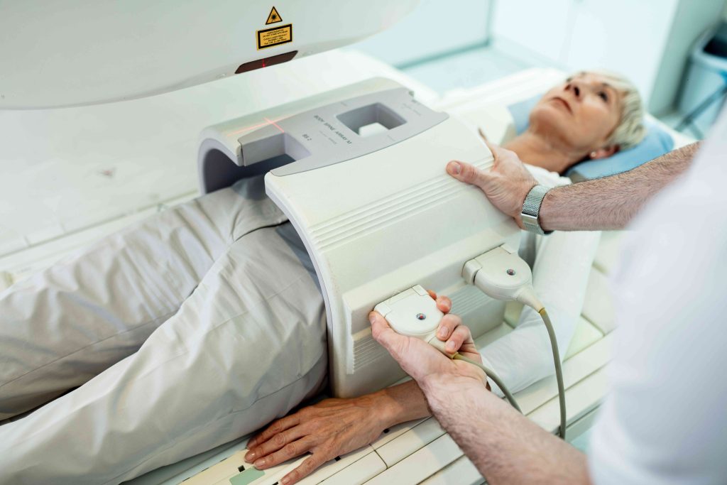
MRI Defecography
MRI defecography uses magnetic fields instead of X-rays to create detailed images of the rectum and pelvic floor, especially soft tissues that X-ray defecography may not clearly show.
The MRI machine generates a strong magnetic field that aligns hydrogen atoms in your body and then captures signals to form images. This test can be done while sitting or lying down.
Being well-prepared for the defecography test can reduce discomfort and confusion and ensure the test goes smoothly. A few things you may consider are:
- Location: The defecography test is done at an independent imaging center or a hospital. A radiologist performs the test following recommendations by a healthcare provider.
- Attire: You need to change into a hospital gown for the test. Avoid wearing metal and electrical objects such as jewelry, hair clips, watches, mobile phones, and tracking devices.
- Food and drink: Consult your healthcare provider to determine if you should avoid eating or drinking before the test. Your provider will often recommend a liquid diet before undergoing the test.
- Medications: Talk to your healthcare provider about any prescriptions and supplements you are taking.
- Items to bring: Bring detailed instructions from your healthcare provider in case your radiologist has any questions.
- Emotional support: If you are stressed or anxious, ask your healthcare provider or radiologist if you can bring a family member or friend for support.
- Cost and insurance: Talk to your insurance company to see if they will cover the cost of the test or if it is your responsibility.
Defecography can take place either using an X-ray or MRI. You may want to talk to your healthcare provider to understand how to prepare and what to expect during the test.
Before the Test
Before your defecography test, your healthcare provider will review your medical history to check for any health conditions, recent surgeries, or implants. They may also give you instructions on eating, drinking, and taking medications before the test. If you have claustrophobia, they might suggest a mild sedative to help you stay comfortable.
To prepare for the test, your provider may perform a bowel-clearing procedure. A contrast agent makes your internal organs visible on the scan; X-ray defecography typically uses a barium paste, while MRI defecography uses gadolinium. The contrast agent is placed into your rectum using a catheter. You sometimes need to drink a contrast solution about an hour before the test to help see your small intestine.
During the Test
You need to change into a hospital gown for the test. The radiologist who performs the test may give you further instructions.
For X-ray defecography, you’ll sit on a special commode that allows X-rays to pass through, with the X-ray tube positioned toward your pelvis. For MRI defecography, you may lie on a moveable examination table with your knees bent inside a traditional MRI machine or sit on an adjustable chair in an open MRI machine placed between two large magnets.
The radiologist will ask you to perform different activities similar to performing a bowel movement, including:
- Squeezing: Tightening the anal sphincter and pelvic floor muscles
- Straining: Pushing as if trying to go but without fully emptying
- Defecating: Emptying the rectum without stopping
The radiologist captures images at each stage to see how your muscles function and help identify any issues. The entire test takes about 30 minutes to an hour to complete.
After the Test
Once the procedure is complete, you can resume your daily activities and normal diet almost immediately.
X-rays and MRIs are relatively safe and do not cause any major side effects. However, a few risks include:
- Ionizing radiation from X-rays can sometimes be harmful, especially for younger people.
- X-rays may not be safe for pregnant people.
- MRIs are generally considered safe for pregnant people but are not recommended, as the fetus may lead the results to be inaccurate.
- In rare cases, you may be allergic to the contrast agent, leading to allergic reactions.
- MRI defecography can cause burns or other harmful reactions if you have metal or electronic implants in your body.
The radiologist will analyze the images and send a signed report to your healthcare provider. Your provider will discuss the report with you and figure out if you have a pelvic floor disorder or condition affecting your rectal function.
If treatment is needed, they can help you find the best options for your case.
Interpretation of Results
The results assess how well different parts of your anorectal area are working. They measure the angle of your rectum, how much your perineum moves, how your pelvic muscles function, the length and opening of your anal canal, and how completely your rectum empties. In people with typical function, results include:
- Anorectal angle: The anorectal angle is formed by a straight line passing through the axis of the anal canal and the other passing through the posterior rectal wall. The anorectal angle is generally 95 degrees with a variation of 65-100 degrees. It needs to get smaller when you contract your rectum and larger when you push stool out.
- Perineal descent: The perineum is the area between the anus and vulva in people assigned female at birth and the anus and scrotum in people assigned male at birth. Perineal descent is when the perineum bulges down from its typical position. In a healthy state, there is no significant perineal descent.
- Puborectalis muscle: This pelvic floor muscle is associated with the lower rectum. It typically relaxes and lengthens when emptying the rectum.
- Anal canal length: The length of the anal canal typically ranges from 2.5 to 4 centimeters.
- Opening of the anal canal: The opening of the canal length is approximately 1.5 centimeters during rectal emptying. There can usually be a difference of up to 3.5 centimeters between emptying and rest.
- Degree of rectal emptying: Calculating rectal emptying requires measuring the areas obtained from images before and after rectal emptying. A degree of 80% or more is considered normal.
Results that differ from these standard functions may indicate that something is affecting your rectal function and/or pelvic floor.
Defecography helps determine how well the pelvic floor muscles and rectum function during a bowel movement. It also can detect underlying disorders such as constipation, anal pain, and pelvic floor dysfunctions.
Your healthcare provider will recommend an X-ray or an MRI defecography, which a radiologist will perform. Once a report is generated, your healthcare provider will discuss the results with you and recommend a treatment plan if needed.